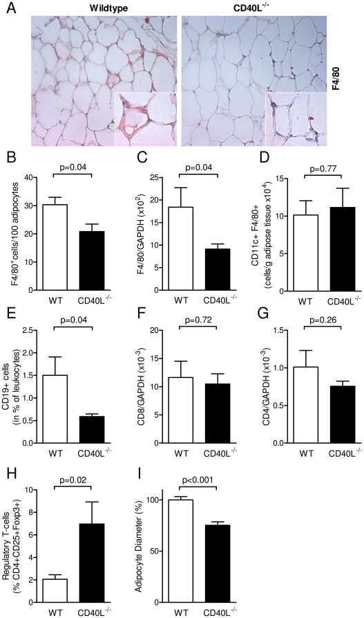
Figure 5. CD40L deficiency attenuates diet-induced adipose tissue inflammation by reduction of immune cell infiltration.
Wildtype (WT) and CD40L−/− mice consumed HFD for 20 weeks. Infiltration of macrophages was quantified by detection of the F4/80 antigen in immunohistochemistry (A, B) and quantitative PCR (C). CD11c-expressing M1-macrophages were determined by flow cytometry (D). B-cells were defined as CD19-positive cells in flow cytometry after enzymatic digestion of adipose tissue and specific anti-CD19 staining (C). Numbers of CD8+ cytotoxic and CD4+ T-helper-cells were quantified by detection of specific mRNA transcripts in whole tissue RNA preparations and normalized to GAPDH (F, G). Regulatory T-cells were defined as CD4+CD25+FoxP3+ cells in flow cytometry (H). Mean adipocyte diameter was calculated in histological sections by image-processing software (I). Data are presented as mean ± SEM of at least 6 animals per group.

