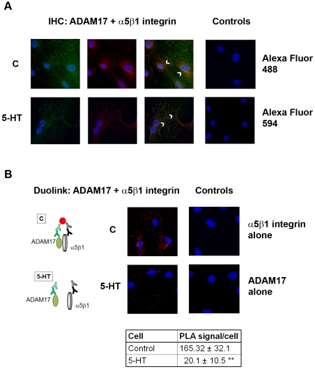
Figure 2. Co-localization of ADAM17 and α5β1 integrin in rat mesangial cells.
Control (C) and 1 µM 5-HT -stimulated mesangial cell were fixed, permeabilized, and (A) co-immunostained using ADAM17 antibody (green) and β1 integrin antibody (red) as indicated in “Material and Methods”. Arrows indicate co-localization of ADAM17 and α5β1 integrin immunopositive areas (yellow). For the negative controls we omitted the primary antibodies and used PBS followed by secondary antibodies. (B) Parallel samples were incubated with oligonucleotide-labeled PLA probes after incubation with primary antibodies. PLA signals as fluorescence dots were imaged and quantified. As negative control we used either ADAM17 or α5β1 integrin antibody alone followed by the oligonucleotide-labeled PLA probes. Cartoon explains binding of the fluorescence detection reagent only to antibodies in close proximity; **p<0.01. Representative examples out of three experiments are shown.

