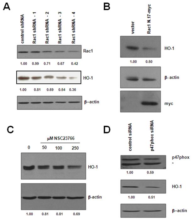
Figure 5. Targeting the NADPH oxidase through Rac1 or p47phox influences HO-1 expression.
(A) Rac1 knockdown affects HO-1 protein level. BaF3-p210 expressing cells were transfected with control or Rac1 specific shRNA and Rac1 and HO-1 protein levels were monitored in total cell lystates by Western blot using Rac1 and HO-1 antibodies, respectively. (B) Rac1 was inhibited by transfection of a dominant-negative Rac1 construct (Rac1N.17-myc). HO-1 and dominant-negative Rac1 expression was evaluated in total cell lysates by Western blot using HO-1 and myc specific antibodies, respectively. (C) BaF3-p210 expressing cells were treated for 48 hours with increasing doses of the Rac1 inhibitor, NSC23766, and HO-1 protein expression monitored by Western blot. (D) BaF3-p210 expressing cells were transfected with control or p47phox specific siRNA and HO-1 protein levels were monitored in total cell lystates by Western blot. Knockdown efficiency was evaluated in the same lysates using p47phox specific antibodies. The * indicates a non-specific cross-reacting band. β-actin was used as a loading control for all Western blots and densitometry was performed using ImageJ software.

