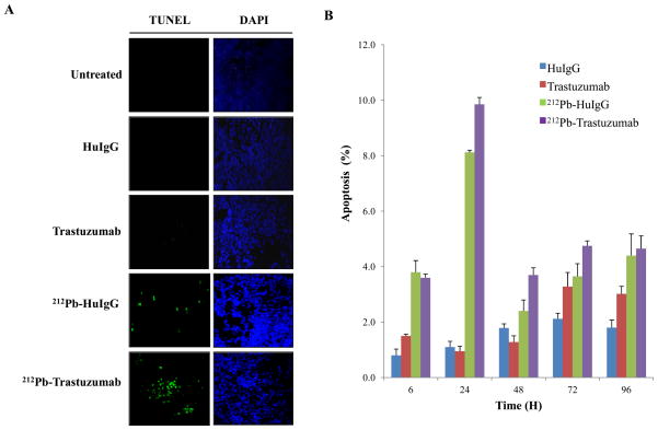
Figure 1. Induction of apoptosis in LS-174T i.p. xenografts following 212Pb-TCMC-trastuzumab.
A. Mice bearing i.p. LS-174T xenografts were treated with 212Pb-TCMC-trastuzumab and the tumors collected over a 120 h period. Additional groups included untreated, HuIgG, and trastuzumab, and 212Pb-TCMC-HuIgG as a non-specific control with the representative fluorescence microscopy images at 24 h. Left, TUNEL staining; Right, DAPI counter staining (40x)
B. Apoptosis induced by 212Pb-TCMC-trastuzumab. Mice bearing i.p. LS-174T xenografts were treated with 212Pb-TCMC-trastuzumab at the indicated times. Paraffin-embedded sections were stained with H&E and the apoptotic nuclei were counted under light microscopy; 500 nuclei were scored per tumor. Results represent the average of a minimum of three replications (± SD).

