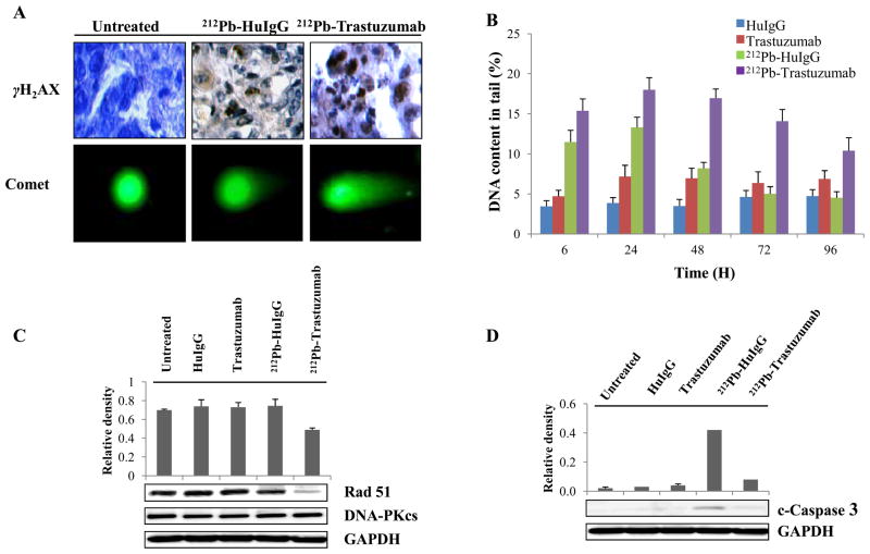
Figure 2. 212Pb-TCMC-trastuzumab-induced DNA damage and repair delay in LS-174T i.p. xenografts.
A. Light and fluorescence microscopy images of untreated, 212Pb-TCMC-HuIgG and 212Pb-TCMC-trastuzumab at 24 h. Top, IHC staining (γH2AX); bottom, neutral comet assay (40x)
B. DNA content in the tail. DNA damage was quantified by % DNA content in the tail using comet assay at the indicated times. Results represent the average of a minimum of three replications (± SD).
C. Down-regulation of Rad51 induced by 212Pb-TCMC-trastuzumab. Immunoblot analysis for Rad51 and DNA-PKcs were performed at the 24 h time point. Rad51 was detected at 37 kDa and DNA-PKcs was detected at 450 kDa. The equivalent protein loading control was GAPDH.
D. Down-regulation of cleaved caspase-3 induced by 212Pb-TCMC-trastuzumab. Immunoblot analysis for cleaved caspase-3 was performed at the 24 h time point. Cleaved caspase-3 was detected at 17 kDa. The equivalent protein loading control was GAPDH.

