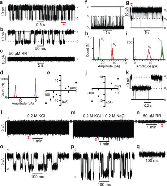
Figure 5. mpiezo1 forms ruthenium red sensitive ion channels.
(a-e) Reconstitution of purified mpiezo1 into asymmetric lipid bilayers. (a) Representative single channel currents at –100 mV. The section of the recordings indicated by the red asterisk is shown in (b) at a 100-fold higher time resolution. (c) After 35 minutes of recording the channel activity shown in (a), injection of 50 μM RR onto the neutral facing compartment blocks mpiezo1 currents. (d) All-event current amplitude histogram of a 6 minute recording; γ = 124 ± 7 pS. The total number of opening events (N) analyzed was 18,424. (e) Single channel current-voltage (IV) relationship, n=6 experiments. (f-k) Reconstitution of purified mpiezo1 into asolectin proteoliposomes. Representative channel currents recorded at –100 mV (f) and +100 mV (g) in presence of 50 μM RR inside the recording pipette. Two open channels are present in the membrane. The segment of the 15 minute recording shown in (g) indicated by the red asterisk is displayed in (k) at a 25-fold higher time resolution. (h,i) All-event current amplitude histograms from a 30 s (h) and 15 min (i) recordings: γ = 110 ± 10 pS (h) and 80 ± 5 pS (i); N was 9,938 events. (j) Single channel I-V relationship, n=8 experiments. (l-q) Representative single channel currents at -100 mV of purified mpiezo1 reconstituted into asymmetric lipid bilayers in symmetric 0.2 M KCl (l), after addition of 0.2 M NaCl (m) and after addition of 50 μM RR (n). Segments indicated by red asterisks in panels l-n are displayed in panels o-q, respectively. C and O denote the closed and open states.

