
Figure 2.
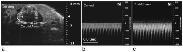
Maternal ethanol exposure (by either intragastric gavage or intraperitoneal injection did not result in a significant change in maternal heart rate or other hemodynamic measures (see results). (a) Sample Doppler image showing angle of insonation and placement of the data acquisition cursor over the maternal common carotid artery. (b and c) show sample sonograms before and after ethanol exposure. Note that the scaling of the ‘Y’ axis (velocity in mm/sec) is different for images b and c, however, there was not a significant difference in maternal carotid artery peak velocity following ethanol exposure. Abbreviations, A; artery. Scale bar (for b and c), 0.6 sec.