Figure 6.

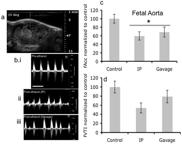
Doppler ultrasound measurement of fetal Aorta. (a) Doppler ultrasound image of fetal Aorta indicating area of analysis. (b) Doppler ultrasound waveform of fetal Aorta before ethanol treatment and after both IP and Gavage routes of administration. (c,d) Blood flow as measured by Acceleration was significantly reduced following maternal ethanol exposure (asterisk indicates statistically significant difference from baseline control), however, VTI, though exhibiting a declining trend, was not significantly decreased. No difference was observed between routes of administration. Scale bar, 0.6 sec.
