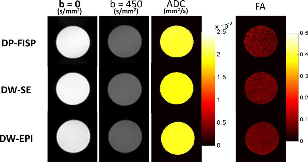
Figure 2.
Representative axial phantom diffusion weighted images, ADC, and FA maps for DP-FISP, DW-SE and DW-EPI methods (b=0 and b=450 s/mm2, six directions). Mean values of these diffusion parameters from five repeated measurements are shown in Table 1.

