
Figure 1.
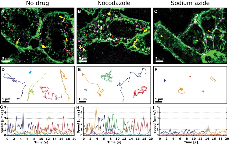
Motion of PV in untreated and nocodazole or sodium azide-treated cells. Confocal images (A–C) of PV (magenta) in live cells labeled with R18 (green; see also Movie S1, Movie S3, and Movie S4). Representative trajectories are overlaid in yellow. Magnified views of these trajectories are shown in D–F, and their instantaneous speed as a function of time are shown in G–I. Cells were pretreated with drug (60 μM nocodazole or 20 μM sodium azide) for 50 min before incubation with Cy5-labeled PV for 10 min at a low multiplicity of infection (MOI = 1 pfu/cell). Just before imaging, cells were briefly incubated with R18, which nonspecifically stained the plasma membrane and allowed the determination of the boundary of the cell and the observation of R18-stained vesicles. The red arrowheads in A and B indicate examples where PV colocalizes with R18-labeled vesicles. Successive points in trajectories are separated by 0.25 s.