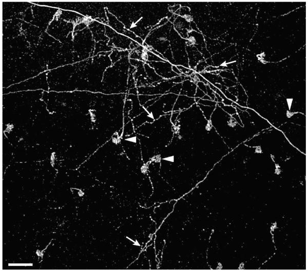
Figure 2. Comparison of wEFs and rEFs.
Fluoro-Ruby labeled terminals shown at the border of the INL and IPL in the retina. The distinctive pericellular nests (arrowheads) can be seen as the terminals of 23 rEFs. In addition, the diffuse arborization of one or more wEFs (arrows) is shown. Unlike the rEFs, wEFs give rise to collaterals and have multiple swellings (likely synaptic boutons) throughout their axonal arborization. Scale Bar is 50 μm.

