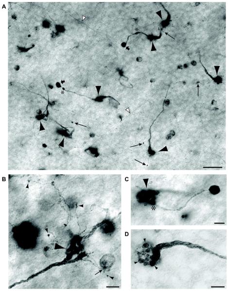
Figure 5. NADPH-diaphorase staining of rEF terminals.
A: NADPH-diaphorase stained flat mount viewed at the INL/IPL boundary. Several rEF fibers give rise to large pericellular nests slightly out of the plane of focus (large arrowheads). Long arrows designate tiny boutons terminating fine “tendrils” arising from the pericellular nest. Another type of secondary synaptic structure, the “ball and chain” comprises a relative thick process, often with swellings (asterisks) terminating in a dense ball (broad arrows). The somata of other diaphorase-positive amacrine cells and target cell axons (white arrowheads) can also be seen in this image. Scale Bar is 20 μm. B: A high magnification image of a single rEF terminal to show the structure of tendrils in greater detail. Tendrils can be seen to branch off the pericellular nest (large arrowhead). These very slender branches terminate in small synaptic boutons on unknown targets in the amacrine cell layer (small arrowheads). One bouton contacts a weakly diaphorase positive amacrine cell (small arrow). Scale Bar is 5 μm. C: A high magnification image of a single rEF terminal to show the structure of a ball and chain in greater detail. A single chain (diameter 1-2 μm) branches off (*) the pericellular nest (arrowhead) and terminates in a large (approx. 5 μm) intensely NADPH-diaphorase positive ball. Scale Bar is 5 μm. D: A pericellular nest at high magnification is seen to comprise about 15, 2 μm diameter, presynaptic structures (arrowheads), giving it the appearance of a bunch of grapes at the end of the swollen efferent fiber. Scale Bar is 5 μm.

