
Figure 3.
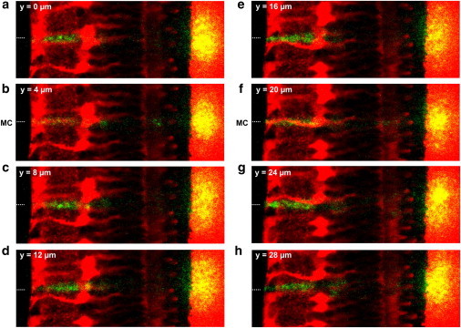
Müller cells bias the transretinal light path. (a–h) Overlay of a fluorescent retinal slice (red) and the scattering of the laser light (green, yellow) applied by a thin glass fiber in front of the retinal surface (position indicated by a dotted line). When the retina was moved in equal steps of 2 μm (every second position is shown) along the optic fiber, the laser scattering in the retina (green) and the scattering of the transmitted light at the membrane (yellow) changed in dependence upon the presence of a Müller cell in the light path. (b and f) If the center of a Müller cell was positioned in front of the fiber core (indicated by MC in front of the retinal surface), the intensity of light scattering in the IPL was reduced. In addition, the transmitted light was confined to a small area and became more intense. (f) These effects were most obvious if a Müller cell was in the focal plane of the objective (indicated by its strong red fluorescence), i.e., at the same z level where the fiber core was placed.