Figure 1.
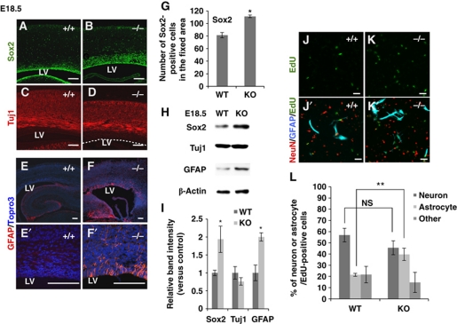
RP58 depletion causes increased astrogenesis both in vivo and in vitro. (A–D) Representative images of coronal sections from E18.5 WT and mutant mice stained for Sox2 (A, B) and Tuj1 (C, D). Scale bars: 0.1 mm. LV, lateral ventricle. The dotted line in (D) indicates the edge of the neocortical VZ (n=3). (E–F′) Representative images of caudal coronal sections from E18.5 WT and mutant mice stained for GFAP. (E′, F′) Show magnified areas in (E, F), respectively. Nuclei were stained with Topro3 (E–F′). Scale bars: 0.1 mm. (G) The number of Sox2-positive cells within a fixed area was counted. (H) Immunoblotting for Sox2, Tuj1, GFAP, and β-actin in cortical homogenates from E18.5 WT and mutant mice. β-Actin was used as an internal control. (I) Quantification of protein expression levels in (H) using Fiji software available online (http://pacific.mpi-cbg.de/wiki/index.php/Fiji) (t-test: *P<0.05, **P<0.01). Error bars indicate the s.d. (n=3). (J–L) Cerebral cortical cells prepared from E16.5 WT or mutant mice were cultured in EdU-containing medium for 12 h and then incubated for 5 days. (J–K′) Cells were stained with antibodies against GFAP (blue) and NeuN (red). Cells incorporating EdU were detected using the alkyl-azide reaction (green). Scale bars: 50 μm. (L) Percentage of neurons, astrocytes, and other cell types within the EdU-labelled progenitor cell population in (J′, K′) (t-test: **P<0.01). Error bars indicate the s.d. (n=7: WT=4, mutant=3).

