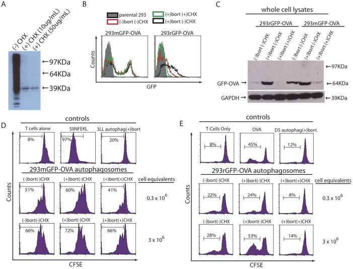
Figure 4.
Inhibition of protein synthesis before proteasome blockade prevents accumulation of short-lived proteins and limits the ability of autophagosomes to stimulate antigen-specific T cells. (A) HEK 293T cells were pulsed with a non-radioactive-labeled methionine while treated with cyclohexamide or vehicle for 4 hours at the indicated concentrations. Cells were harvested and a western blot was performed to detect any labeled proteins during the 4-hour pulse period. (B and C) HEK 293T cells expressing a stable GFP-OVA fusion protein (mGFP-OVA) or a short-lived GFP-OVA fusion protein (rGFP-OVA) were treated with bortezomib for 4–24 hours or pretreated with cyclohexamide for 4 hours before addition of bortezomib. (B) Expression of GFP-OVA 4 hours after addition of bortezomib measured by flow cytometery. Filled histogram (parental HEK 293T cells), red line (untreated cells), black line (cells treated bortezomib) and green line (cells pretreated with cyclohexamide for 4 hours followed with bortezomib). (C) Expression of GFP-OVA 24 hours after addition of bortezomib or CHX and bortezomib measured by western blot with anti-GFP in total cell lysates from treated cells. (D and E) Autophagosomes were isolated from 293rGFP-OVA or 293mGFP-OVA cells treated as in (C). Non-specific control autophagosomes were isolated from (D) 3LL carcinoma or (E) D5 melanoma. Autophagosomes were pulsed onto APCs and used to stimulate naïve antigen-specific OT-1 T cells. Proliferation was measured by CFSE dilution. (D) 293mGFP-OVA derived autophagosomes n=2 experiments. (E) 293rGFP-OVA derived autophagosomes n=3 experiments.

