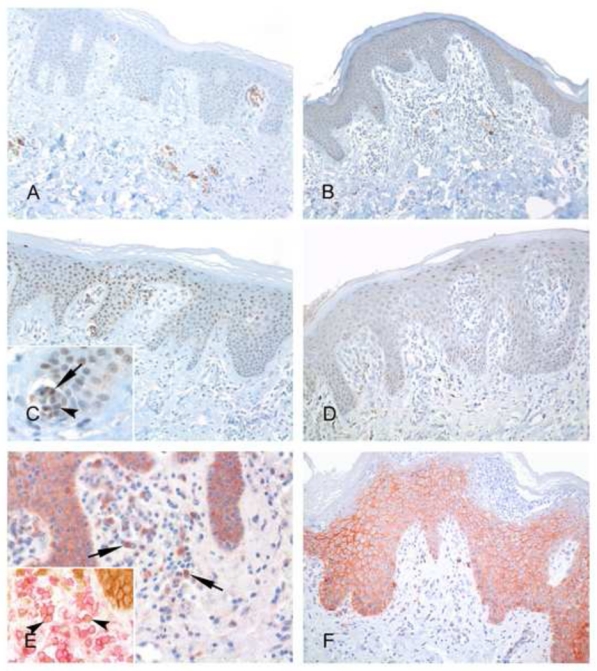
FIGURE 2. Representative immunohistochemical results for CD30, beta-catenin and JunB expression in erythrodermic CTCL and inflammatory dermatoses.

A, C and E show examples of CTCL, while B, D, F are from the EID group. A, Clusters of CD30+ atypical lymphocytes are present around dermal vessels in this Sézary syndrome skin sample. B, Presence of scattered CD30+ lymphocytes is evidenced in this example of erythrodermic psoriasis. C, A subset of atypical lymphocytes within Pautrier’s microabcesses show a positive nuclear staining for JunB (inset, arrows), with a similar staining in the surrounding keratinocytes, used as a positive control. D, In this EID sample, no JunB positive lymphocytes are seen, whereas a distinctive staining of keratinocytes nuclei is evidenced. E, Few β-catenin positive lymphocytes can be demonstrated in this Sézary syndrome sample (arrows), whereas keratinocytes expressing this antigen at their membrane can be seen in the overlying epidermis, used as a positive control. Most β-catenin positive (brown) lymphocytes are T-cells, as shown by double immunostaining experiments with CD3 (red) (inset, arrowheads). F, In this pustular erythrodermic psoriasis, no β-catenin positive lymphocytes are seen.
