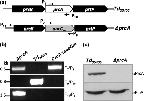
Fig 3.
Isolation and characterization of ΔprcA mutant. (a) Illustration of the prcA locus in Td35405 (wild type) and the aacCm cassette in the ΔprcA mutant. (b) PCR analysis showing that the prcA gene was deleted and replaced by aacCm in the ΔprcA mutant. The primers used for PCR analysis are labeled in panel a. (c) Western blot analysis of the ΔprcA mutant. Similar amounts of whole-cell lysates from the wild type and the mutant were analyzed by SDS-PAGE and then probed with a specific PrcA antibody (αPrcA, anti-PrcA antibody). Immunoblots were developed using horseradish peroxidase secondary antibody with an enhanced chemiluminescence (ECL) luminol assay as previously described (2, 3).

