Fig 1.
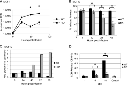
M. marinum pathogenesis within A. castellanii requires the ESX-1 secretion system. (A) M. marinum replicates and persists within A. castellanii, while the M. marinum strain lacking ESX-1 is attenuated. Amoebae were infected (MOI of 1) and treated with gentamicin (150 μg/ml). Amoebae were lysed and bacteria were plated for CFU at the indicated times. Error bars represent standard deviations. Asterisks represent statistically significant differences based on an unpaired tailed Student t test (P < 0.05) (48 hpi, P = 0.035; 72 hpi, P = 0.002). (B) Infection by M. marinum results in a stable percentage of infected amoebae, while the population of infected amoebae decreased over time in the absence of the ESX-1 system. A. castellanii was infected with M. marinum and ΔRD1 M. marinum expressing DsRed at an MOI of 10. Monolayers were imaged using a 20× objective on a Zeiss AxioObserver microscope. The percentage of infected amoebae was established after counting >100 cells in each of five different fields at the indicated time points (12 h, P = 0.005; 24 h, P = 0.010; 48 h, P = 6.547 × 10⁻⁵). (C) Infection of A. castellanii results in a relatively constant number of amoebae over time, while infection by M. marinum lacking ESX-1 leads to amoeba growth. At each time point following infection, the amoebae were counted and the number was normalized to the initial number of amoebae plated. See Fig. S3 in the supplemental material for amoeba counts. (D) Infection with M. marinum results in amoeba lysis in an ESX-1-dependent manner. MOI of 5, P = 0.009; MOI of 10, P = 0.001. The control is uninfected amoebae after 72 h of incubation.

