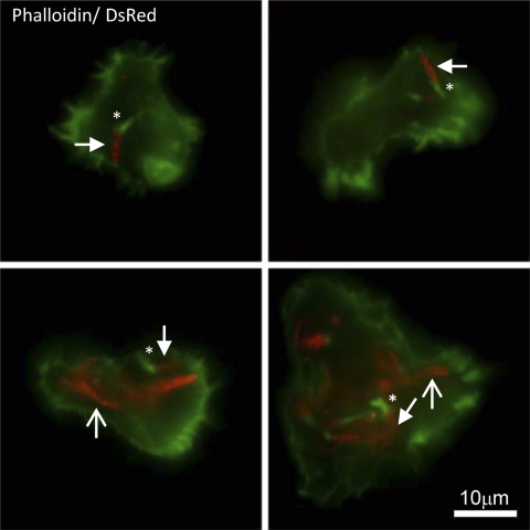
Fig 2.
Wild-type M. marinum forms actin tails in A. castellanii. Florescence microscopy demonstrating actin tail formation by M. marinum at 22 hpi, MOI of 5. M. marinum is expressing DsRed, and Alexa 488 phalloidin is green. Four representative images are shown. The scale bar is 10 μm. Images were acquired with an Evolution QEi charge-coupled device (CCD) (Media Cybernetics) on a Nikon Eclipse TE300 microscope (60× objective) using IPLab software (Scanalytics). Examples of bacteria bearing tails are indicated with filled arrows. Tails are indicated with asterisks. Examples of bacteria without tails are indicated with open arrows.

