Figure.
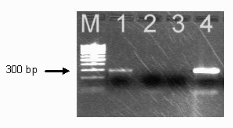
Polymerase chain reaction amplification of Anaplasma phagocytophilum DNA from the patient's acute-phase blood sample. Amplified DNA was separated by electrophoresis through the 2% agarose gel stained with ethidium bromide. Lane 1, patient sample (note the presence of the band at ≈293 bp); lane 2, negative sample; lane 3, negative control (no-DNA template control); lane 4 positive control (DNA extracted from the cultured isolate of A. phagocytophilum). Lane M represents a 100-bp DNA ladder for estimation of molecular sizes.

