Figure 3.
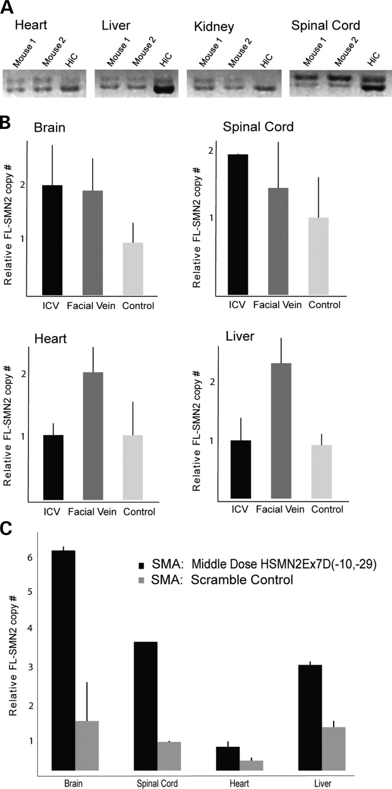
Central versus peripheral SMN2 splice modulation. (A) RT–PCR systemic analysis of full-length SMN2 after P0 MO ICV injection. There is no increased SMN2 exon 7 incorporation in visceral structures (the heart, liver, kidney) when compared with spinal cord (n= 2). (B) ddPCR (full-length SMN2 relative to cyclophilin) of the brain, spinal cord, heart and liver after P0 MO ICV (54 µg) or P0 FV (50 µg/g) injection in Smn+/−; SMN2+/+; Δ7SMN+/+ mice. Results per organ are relative to scramble-injected control, which has been set at 1 on the y-axis. There is increased CNS splice modulation after both ICV and peripheral dosing. There is no increased splicing in the heart and liver after ICV injection, suggesting limited translocation across the CSF–blood barrier or rapid systemic degradation. (C) ddPCR (FL-SMN2 relative to cyclophilin) of the brain, spinal cord, heart and liver after P0 MO ICV (54 µg) injection in Smn−/−; SMN2+/+; Δ7SMN+/+ mice; SMA scramble-injected control per each organ. Results are displayed as absolute relative ratios on the y-axis. There are large increases in CNS splice modulation, and a more modest increase in liver full-length SMN2.

