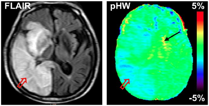
Figure 5.

Conventional and pHW MR images of a patient with stroke at 5 days post-onset. The hyperintense stroke area identified on the FLAIR image (T2W with CSF suppressed) by the red arrow is hypointense on the pHW image. (Reproduced, with permission, from Zhao X, et al. Magn Reson Med. 2011;65:doi: 10.1002/mrm.22891.)
