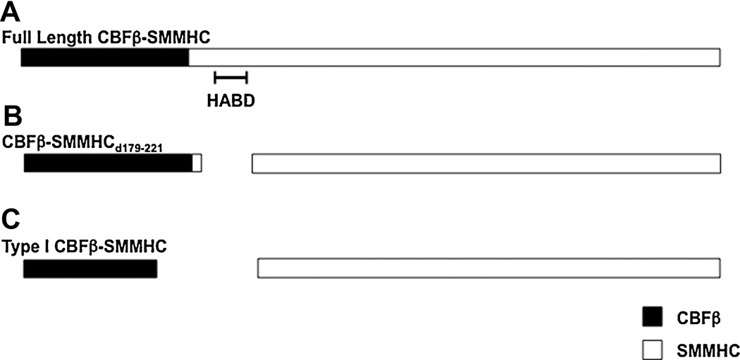
Figure 1. Diagrammatic representation of CBFβ-SMMHC variants.
Schematic of (A) full length CBFβ-SMMHC, (B) the CBFβ-SMMHCd179-221 deletion mutant, and (C) the Type I CBFβ-SMMHC fusion. The CBFβ and SMMHC are represented as black and white boxes, respectively. The high affinity binding domain (HABD) is indicated.

