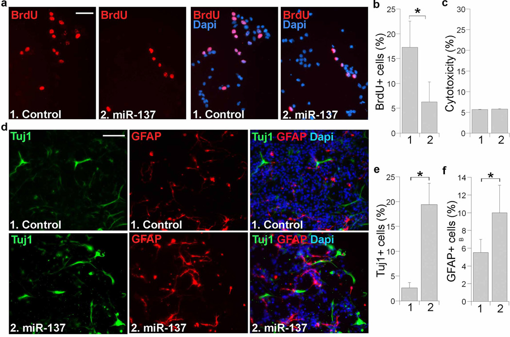
Fig. 1. Overexpression of miR-137 regulates neural stem cell proliferation and differentiation.
a. Cell proliferation in miR-137-transfected embryonic neural stem cells as revealed by BrdU labeling (red). Right panels show BrdU staining along with Dapi counter staining. Scale bar, 50 µm. b. Quantification of BrdU-positive (BrdU+) cells in miR-137-treated neural stem cells with s.d. indicated by error bars. * p<0.001 by Student’s t-test. n=7. c. Minimal cytotoxicity in miR-137-transfected neural stem cells, as revealed by LDH assays for cytotoxicity. n=4. d. Overexpression of miR-137 promotes neural differentiation. Control RNA or miR-137-transfected cells were induced into differentiation for 5 days and immunostained with a Tuj1-specific antibody (green) or a GFAP-specific antibody (red). Nuclear Dapi staining is shown in blue. Scale bar, 100 µm. e. Quantification of Tuj1-positive (Tuj1+) cells in control and miR-137-treated neural stem cells. n=3. f. Quantification of GFAP-positive (GFAP+) cells. n=3. For both sections e and f, error bars are s.d. of the mean. *p<0.005 (e), *p<0.1 (f) by Student′s t-test. About 4,000 cells were quantified for sections b, e and f, respectively.

