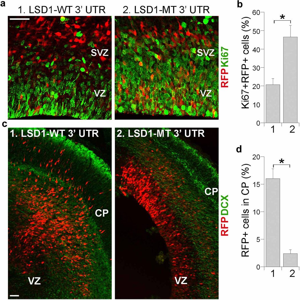
Fig. 5. Mutation of LSD1 3’ UTR rescues miR-137-induced proliferative defect and precocious migration.
a. In utero electroporation of a LSD1 vector with its wild type 3’ UTR (LSD1-WT 3’ UTR, 1) or mutant 3’ UTR (LSD1-MT 3’ UTR, 2) along with miR-137 into E13.5 mouse brains. The miR-137 + LSD1-MT 3’ UTR treatment, but not miR-137 + LSD1 WT 3’ UTR treatment increased cell proliferation in the ventricular zone (VZ) and subventricular zone (SVZ) of electroporated brains. The electroporated cells were RFP+. Proliferating cells were labeled by Ki67. Scale bar, 50 µm. b. Percent of Ki67-positive cells out of RFP-positive cells (Ki67+RFP+/RFP+) in miR-137 + LSD1-WT 3’ UTR (1) and miR-137 + LSD1-MT 3’ UTR (2)-electroporated brains. s.d. is indicated by error bars. * p<0.005 by Student′s t-test. n=3. c. Electroporation of miR-137 + LSD1-MT 3’ UTR led to significantly less outward cell migration. The electroporated cells were shown red due to the expression of RFP. Merged images of RFP and double cortin (DCX) staining were shown. CP: cortical plate. Scale bar, 100 µm. d. Quantification of RFP-positive (RFP+) cells that were migrated to the CP. Error bars are s.d. of the mean. *p<0.001 by Student’s t-test. n=3.

