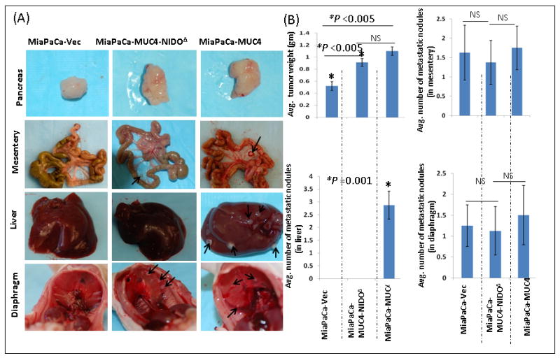
Figure 3. Macroscopic examinations of orthotopic pancreatic tumors and organs with metastatic lesions.

(A). Macroscopic appearance of primary pancreatic tumors from animals implanted with MiaPaCa-MUC4, MiaPaCa-MUC4-NIDOΔ and MiaPaCa-Vect cells. There was no difference in the gross appearance of tumors derived from MiaPaCa-MUC4 and MiaPaCa-MUC4-NIDOΔ groups. However, tumors of the MiaPaCa-Vect group were smaller than the other two groups. Further, metastasis to organs such as the diaphragm and mesentery was same in all the three groups excluding liver metastasis, which was observed only in animals injected with MiaPaCa-MUC4 cells. (B) Quantitative analysis showed a significantly smaller tumor generated from MiaPaCa-Vect cells than MiaPaCa-MUC4 and MiaPaCa-MUC4-NIDOΔ. Further analysis of metastatic lesions among the three groups showed no significant difference in the number of metastatic nodules that were detected in mesentery and the diaphragm. However, there was a significant difference (p=0.001) in the number of metastatic nodules present in the liver.
