
Figure 4. Microscopic examinations of orthotopic pancreatic tumors and organs with metastasis lesions.
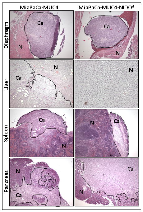
Histological appearance of pancreas, spleen, liver and diaphragm showing prominent metastasis in the mice implanted with MiaPaCa-MUC4 or MiaPaCa-MUC4-NIDOΔ cells. Between these two groups, there was no gross difference in the local tissue invasion pattern. Normal (N) and cancer cells (Ca) in different organs are separately indicated.