
Figure 5. MUC4 NIDO domain interactions with Fibulin-2 in vitro, and colocalisation in pancreatic tissue.
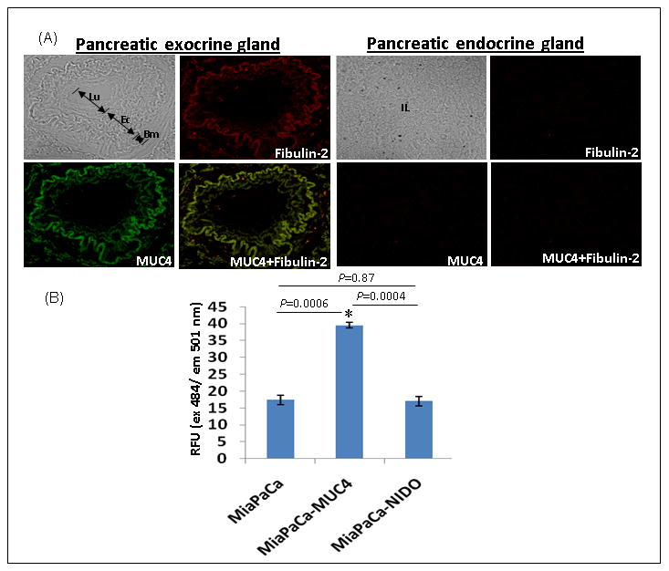
(A) Immunohistofluorescence analysis was carried out on formalin-fixed and paraffin-embedded pancreatic tumor tissue sections after rehydration and antigen-retrieval. Digital merging of the immunostained tissue section clearly showed the colocalization of MUC4 (green) and fibulin-2 (red) in an adenocarcinoma lesion. Absence of MUC4 and fibulin-2 staining in the endocrine glands of the same tissue section acted as a negative control. (B) Binding Assay Cells (25 × 103) were seeded in 96-well plate coated with fibulin-2. Wells coated with bovine serum albumin and poly-L-lysine served as negative and positive controls, respectively. A total of 100 μl of the cell suspension was seeded in triplicates onto the fibulin-2-coated 96 well plates and incubated for 1 h at 37°C. Next the adhered cells were labeled with DIO dye for 1 h at 37°C. The fluorescence of the samples was measured by using the fluorescence plate reader at an excitation wavelength of 484 nm and an emission wavelength of 501 nm. The fluorescence intensity obtained in the negative control (BSA) was subtracted from the values from fibulin-2 coated plates. A relative fluorescence (RFU) was calculated for all treatments with respect to the intensity value obtained with the positive control (poly-L-lysine-coated plates). The significance of each binding assay was evaluated using the t test assuming unequal variances. P-values lower than 0.05 were considered statistically significant.