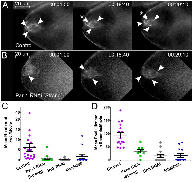
Figure 3. Myo-II Forms Localized Foci in a Par-1-dependent Manner.
(A–B) Frames from time-lapse control (A) and Par-1 RNAi Strong (B) movies (Movie S4) showing Sqh:GFP localization in border cells before detachment at the indicated times (hr:min:s). Transiently enriched foci (arrowheads) were observed throughout the movies. A stable focus is indicated with an asterisk in (A). Par-1 RNAi border cells have fewer foci and Sqh:GFP is generally more diffuse than control. Scale bar, 20 μm. (C–D) Quantification of mean Sqh:GFP foci number (C) and lifetime (D) in movies of the indicated genotypes. Each point represents the average of individual foci measured within a single movie. N ≥ 9 movies for each genotype; P ≤ 0.0001 (unpaired two-tailed t test). Error bars represent SEM. See Supplemental Experimental Procedures for details on analyses.

