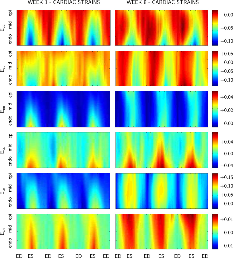
Figure 5.
Spatio-temporal evolution of elastic strains in the lateral left ventricular wall at weeks 1 and 8, time-aligned and averaged over n=11 animals. Components of the Green Lagrange strains are displayed across the ventricular wall for three consecutive heartbeats. C circumferential, R radial, and L longitudinal direction. ED end diastole, EIR end isovolumetric relaxation, ES end systole, and EIC end isovolumetric contraction.

