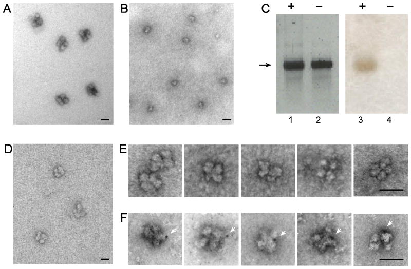
Fig. 3.
Reconstitution of supraspliceosomes from native spliceosomes and Nanogold-tagged pre-mRNA. Native spliceosomes were prepared from supraspliceosomes by specific cleavage of the pre-mRNA as described (Azubel et al., 2004). For reconstitution of supraspliceosomes native spliceosomes were incubated with β-globin pre-mRNA (untagged or tagged with Nanogold) as described (Azubel et al., 2006). (A, B, D) Visualization by EM of a field of supraspliceosomes (A); Native spliceosomes (B); and reconstituted supraspliceosomes (D), negatively stained with 1% uranyl acetate. (C) Gel electrophoresis (2% agarose) analysis of untagged (lanes 2 and 4) and Nanogold-tagged β-globin pre-mRNA (lanes 1 and 3), used for the reconstruction experiment. Left panel, staining with ethidium bromide; right panel, staining of the blotted gel by silver enhancement. Galleries of images of reconstituted supraspliceosomes visualized by EM after staining with 1% uranyl acetate and gold enhancement. (E) Gallery of supraspliceosomes reconstituted from native spliceosomes and untagged β-globin pre-mRNA. (F) Gallery of supraspliceosomes reconstituted from native spliceosomes and Nanogold-tagged β-globin pre-mRNA. White arrowheads point to gold-enhanced Nanogold. Bar, 50 nm.

