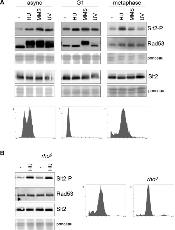
Figure 5.
Analysis of the cell cycle dependent activation of Slt2 by genotoxic stress. A) Exponentially growing cells of the wild type strain (W303-1a) were arrested in G1 by incubation in the presence of 5 μg/mL α-factor for 2 hours. Exponentially growing cells of the GAL1:CDC20 strain (JCY1645) were arrested in metaphase by incubation in YPD medium for 3 hours. Cell cycle arrest was confirmed by cell morphology (more than 95% of unbudded cells or more than 90% of large budded cells respectively) and analysis of DNA content by flow cytometry (lower panels). Once arrested, cells were incubated for 60 min in the absence or presence of 200 mM hydroxyurea or 0,04% MMS, or were exposed to UV radiation (50 J/m2), while maintaing cell cycle arrest with α-factor or glucose. In the case of α-factor arrested cells, 600 mM HU, 0.12% MMS and 150 J/m2 UV radiation was used. The level of phosphorylated Slt2, total Slt2 protein and the chekpoint kinase Rad53 was determined by western analysis. The ponceau staining of the membranes are shown as loading control. B) Slt2 activation by HU in metaphase arrested cells of the GAL1:CDC20 strain (JCY1645) and a rho0 strain derived from it were analysed as described in A.

