
Figure 1.
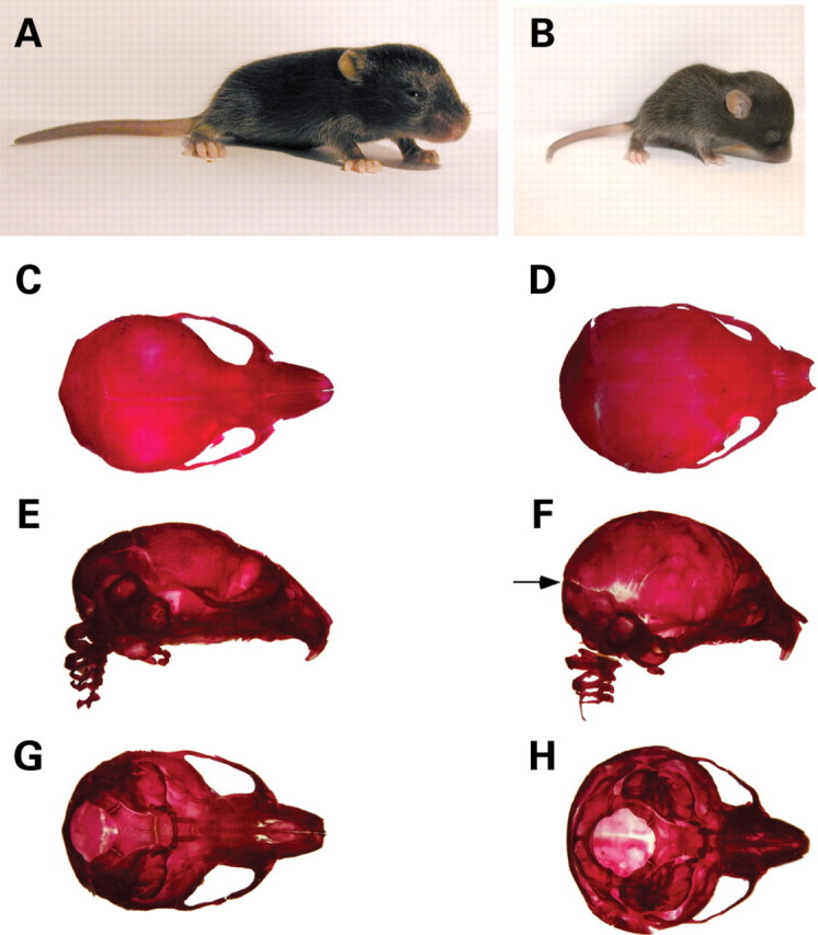
Hydrocephalus in Wnt1-Cre/+; Hdhflox/− mutants. (A and B) Appearance of WT (A) and mutant (B) mice at P13. Note that the mutant mouse (B) is small, runted and displays a domed cranium. (C–H) Skeletal head preparations of WT (C, E, G) and mutant (D, F, H) mice at P10. (C, D) Dorsal view; (E, F) lateral view and (G, H) ventral view. For both WT and mutant, the lower jaw has been removed prior to photographic documentation. Note the enlarged domed cranium in the mutant. Although all skeletal bones appear well formed, suture fusion defects are apparent (arrow in F).