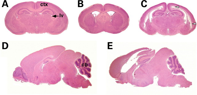
Figure 2.
Ventricular dilation and tissue loss in Wnt1-Cre/+; Hdhflox/− mutant mice. (A–C) H&E histological examinations of coronal sections of P4 WT (A) and mutant (B, C) brains. Extensive dilation of the lateral ventricles (lv) is evident in the mutants at P4 (B), and tissue loss in the cortex (ctx) is observed in more caudal sections (arrowheads in C). (D and E) H&E histological examinations of sagital sections of P14 WT (D) and mutant (E) brains. Note that the progressive hydrocephalus leads to secondary compression of the cerebellum (cb) and more pronounced tissue loss in the mutant brain (E).

