
Figure 3.
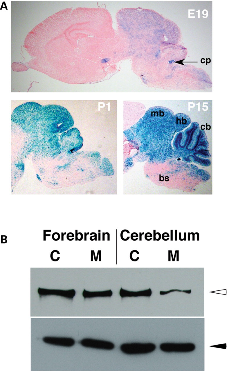
Pattern of Wnt1-Cre-mediated recombination. (A) X-Gal staining of sagital sections of E19, P1 and P15 Wnt1-Cre/+; R26R/+ brains. Note that in all cases, X-Gal staining (blue color) is restricted to the midbrain (mb), hindbrain (hb) and cerebellum (cb). Sparse and scattered staining is observed in the brain stem (bs) and almost no staining is present in the forebrain. cp=choroid plexus. (B) Western analyses of total protein lysates from forebrain (lanes 1 and 2) and cerebellum (lanes 3 and 4) of control (C) Hdhflox/− (lanes 1 and 3) and mutant (M) Wnt1-Cre/+; Hdhflox/− (lanes 2 and 4) mice. Upper panel shows detection of htt (open arrowhead) with the monoclonal anti-htt antibody 2166, and lower panel shows anti-beta-tubulin staining (filled arrowhead), for loading control.