
Figure 4.
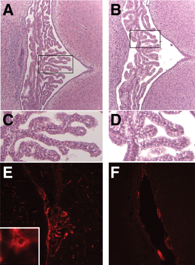
Abnormal choroid plexus in Wnt1-Cre/+; Hdhflox/− mice. (A and B) H&E staining of coronal sections through the fourth ventricle of WT (A) and Wnt1-Cre/+; Hdhflox/− (B) brains at P4. Note that the overall morphology of the choroid plexus appears normal in mutants. (C and D) High magnification of the areas marked by the boxes in (A) and (B), respectively. Note that although the structure of mutant cp (D) is similar to controls (C), a large fraction of the epithelial cells in the mutants display enlarged cytoplasmic volume. (E and F) Immunohistochemistry for htt in the fourth ventricle choroid plexus of P9 WT (E) and Wnt1-Cre/+; Hdhflox/− (F) brains. Note that htt is expressed in choroid plexus epithelial cells of WT mice (see high magnification, inset in E), and that expression is absent in the mutants.