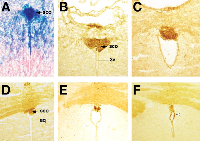
Figure 6.
SCO-spondin expression in Wnt1-Cre/+; Hdhflox/− mutant mice. (A) X-Gal staining of coronal brain sections from Wnt1-Cre/+; R26R/+ mice confirms extensive cre-mediated recombination in cells forming the SCO (blue staining). In addition, X-Gal staining is also observed in the dorsal population of ependymal cells of the aqueduct. (B–F) Immunohistochemistry for SCO-spondin using anti-RF antibody at P18. (B and D) WT and (C, E, F) Wnt1-Cre/+; Hdhflox/− brain sections. Note that progressive hydrocephalus leads to significant dilation of the third ventricle (3v) and aqueduct (aq) in mutant mice (C and E). (B and C) Expression of SCO-spondin is normal in the rostral portion of the SCO in mutants (C) compared to control mice (B). Note that at more caudal levels, RF immunoreactivity is discontinuous in the mutant SCO (E), that SCO is absent in further caudal sections (F), while ectopic RF immunoreactivity is observed in the dorsal ependymal layer of the aqueduct (open arrowhead in F), in a pattern corresponding to the X-Gal staining seen in (A).

