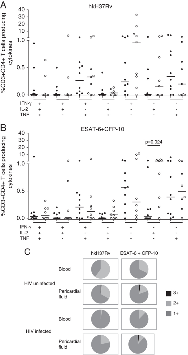
Figure 5.

Polyfunctional CD4+ T cells are present at the disease site. CD3+CD4+ T cells were assessed for the expression of various combinations of IFN-γ, IL-2 and TNF in response to (A) hkH37Rv and (B) ESAT-6+CFP-10 stimulation, in the pericardial fluid of HIV-1-uninfected (closed circles) and HIV-1-infected (open circles) patients. (C) Pie-charts illustrate the overall proportional contribution of cytokine expressing cells to the antigen-specific response in blood and pericardial fluid. 1+: any one cytokine, 2+: any two cytokines, 3+: all three cytokines. IL-2 single-positive cells were proportionally expanded in the HIV-1-infected pericardial fluid compared with the uninfected in response to ESAT-6+CFP-10 (B, p=0.024, Mann–Whitney U test).
