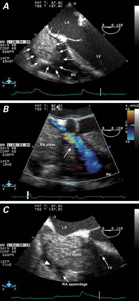
Fig. 1 Transesophageal echocardiography in a patient with superior vena cava syndrome shows A) a mass filling the entire right atrium (RA) and protruding through the tricuspid valve (4-chamber view); B) restricted venous return (arrow) around the RA mass into the right ventricle (RV) (color-flow Doppler), and C) attachment by pedicle (arrowhead) of the RA mass to the RA free wall. LA = left atrium; LV = left ventricle; TV = tricuspid valve

An official website of the United States government
Here's how you know
Official websites use .gov
A
.gov website belongs to an official
government organization in the United States.
Secure .gov websites use HTTPS
A lock (
) or https:// means you've safely
connected to the .gov website. Share sensitive
information only on official, secure websites.
