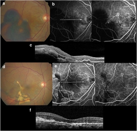
Figure 4.
Images from the right eye of a 63-year-old man with polypoidal choroidal vasculopathy treated with ranibizumab injection. (a) Fundus photograph obtained from at the initial visit showing diffuse subretinal hemorrhage; BCVA was 20/100. (b) FA and ICGA showing a large branching vascular network that terminates with multiple polypoidal lesions. (c) Sectional image obtained with OCT (with the arrow seen on FA) showing protrusions of RPE, which is reflective of the pigment epithelial detachment. (d) The patient was treated intravitreal ranibizumab injections (total injections; four times). Fundus photograph obtained at 12 months after initial visit showing no more subretinal hemorrhage; BCVA was 20/30. (e) FA and ICGA showing regression of the polyps. (f) Sectional image obtained with OCT (with the arrow seen on FA) showing much stabilized elevation of the RPE and no more subretinal hemorrhage.

