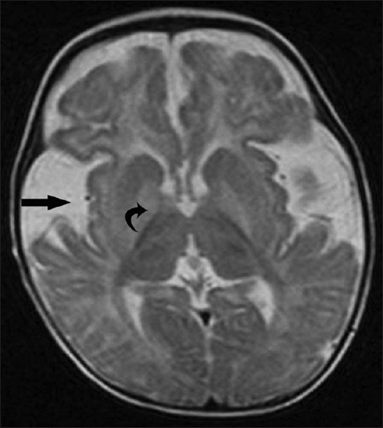
Figure 1.

Axial T2W magnetic resonance image reveals fronto-temporal atrophy, dilated sylvian fissures with open opercula (straight arrow), diffuse white matter signal abnormality and bilateral high signal in the basal ganglia (curved arrow). Widening of the sylvian fissure gives the characteristic “bat-wing” appearance
