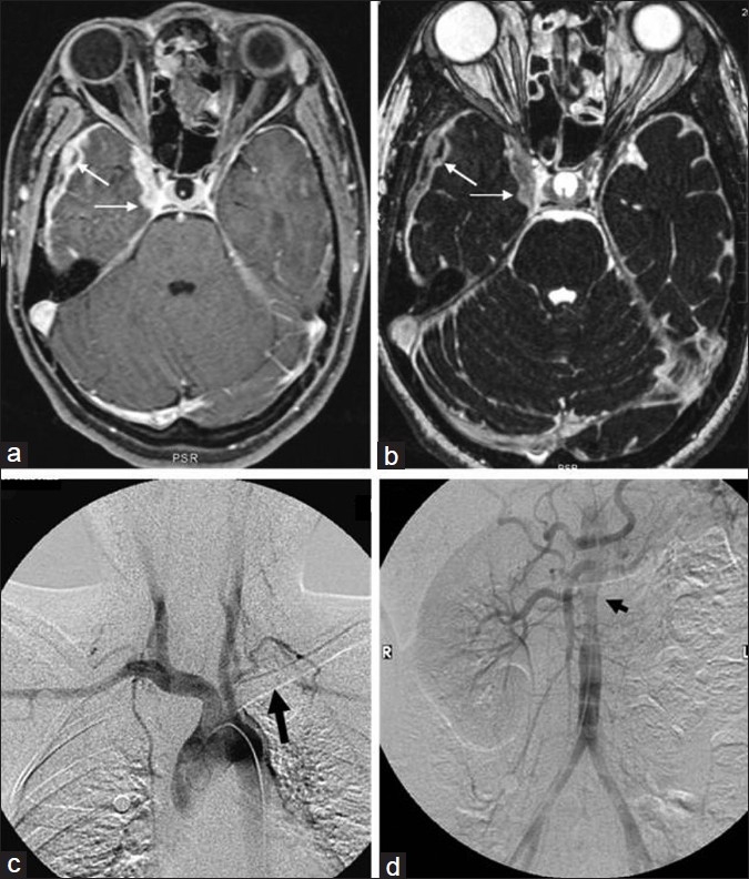
Figure 1.

Axial contrast-enhanced T1-weighted fat saturated image shows thickened enhancing pachymeninges in the right middle cranial fossa and paracavernous region (a-thin arrows). Thickened pachymeninges can also be seen on T2-weighted constructive interference at steady state (CISS) images also (b-thin arrows). Digital subtraction angiography (DSA) of the aortic arch (c) and abdominal aorta (d) shows the left subclavian occlusion (thick arrow) and the non-visualization of the left renal artery with narrowing of the juxta renal abdominal aorta (arrow head), respectively
