Figure 2.

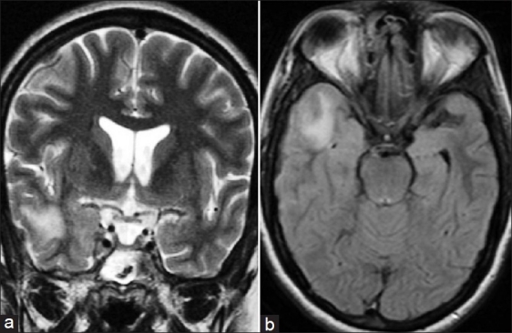
Coronal T2-weighted image (a) and axial Fluid Attenuated Inversion Recovery (FLAIR) sequence (b, shows subcortical white matter hyperintensity in right temporal lobe extending in to the overlying cortex

Coronal T2-weighted image (a) and axial Fluid Attenuated Inversion Recovery (FLAIR) sequence (b, shows subcortical white matter hyperintensity in right temporal lobe extending in to the overlying cortex