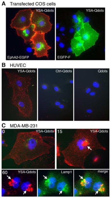
Figure 2. The YSA peptide targets cells expressing EphA2.
(A) The YSA peptide targets quantum dots to EphA2 on the cell surface. COS cells transfected with either the extracellular and transmembrane portions of EphA2 fused to enhanced green fluorescent protein (EphA2-EGFP) or membrane targeted EGFP-F were incubated with YSA bound to red fluorescent quantum dots (YSA-Qdots). YSA-Qdots only bind to cells transfected with EphA2-EGFP. EGFP fluorescence is green; nuclei are stained in blue with DAPI. (B) YSA targets quantum dots to endogenous EphA2 on the cell surface. HUVE cells were incubated at 4ºC with YSA conjugated Qdots (YSA-Qdots), an unrelated 12-mer control peptide that does not bind to EphA2 also conjugated to Qdots (Ctrl-Qdots), or unconjugated Qdots (Qdots). Only quantum dots conjugated to the YSA peptide bind to the HUVE cells. (C) YSA targets Qdots to lysosomes. MDA-MB-231 cells, which express high levels of endogenous EphA2, were incubated at 4ºC with YSA-Qdots followed by incubation at 37 ºC for 0, 15, and 60 min. YSA-Qdots are seen on the cell surface at 0 min but become concentrated in structures near the nucleus at 15 and 60 min (arrow). Double labeling with the lysosomal marker Lamp1 (green) shows colocalization of the quantum dots in lysosomes at 60 min (arrows).

