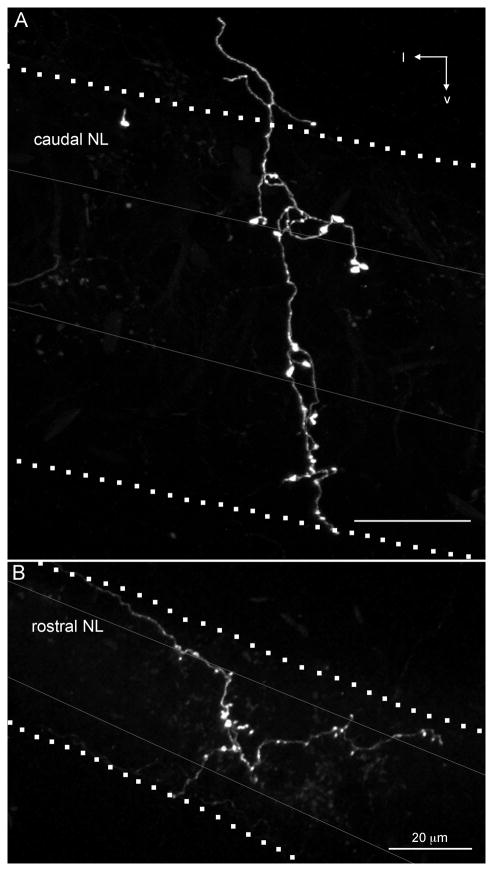
Fig. 10.
SON axonal branches ramifying in high-CF and low-CF regions in NL. AlexaFluor 568 dextran amine labeled SON axons (white) extending across all laminae of NL. In both examples, the axon branch enters NL from the dorsolateral brainstem; however, we observed axons entering NL from both the dorsal and ventral sides. Dotted lines indicate the boundaries of NL, gray lines indicate boundaries of cell body lamina within NL. A:Rostral, high-CF region of NL innervated by an SON axonal arbor. Image is a maximum intensity projection through a 200-μm-thick optical stack. B: Caudal, low-CF region of NL innervated by an SON axonal arbor. Image is a maximum intensity projection through a 300-μm-thick optical stack. l, Lateral; v, ventral. Scale bars = 20 μm.

