Figure 1.

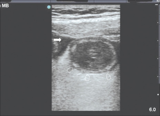
Sonographic section of the central abdomen using a linear probe showing a dilated small bowel (arrow heads) with thickened mucosa and free intraperitoneal fluid (arrow). At laparotomy a segment of the small bowel was gangrenous

Sonographic section of the central abdomen using a linear probe showing a dilated small bowel (arrow heads) with thickened mucosa and free intraperitoneal fluid (arrow). At laparotomy a segment of the small bowel was gangrenous