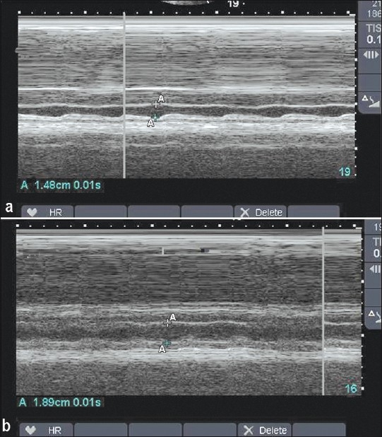
Figure 3.

(a) M-mode of the inferior vena cave before and after (b) resuscitation of the patient shown in Figure 2. Notice the obvious variation in the IVC diameter before resuscitation and the increased diameter of the IVC and less variation in the diameter after resuscitation
