Figure 2.
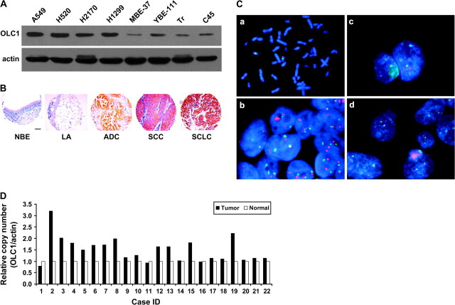
Overexpression and amplification of overexpressed in lung cancer 1 (OLC1) in lung cancer tissues and cell lines. A) Immunoblot of OLC1 in lung cancer cell lines (A549, H520, H2170, and H1299) and immortalized bronchial epithelial cell lines (MBE, YBE, Tr, and C45) using a rabbit polyclonal anti-OLC1 antibody. Blots were probed with mouse monoclonal anti–β-actin antibody as a control for loading and transfer. A representative blot from three independent experiments is shown. B) Representative examples of tissue microarray–based immunohistochemical staining of OLC1 in human lung cancer and normal lung tissues using a rabbit polyclonal anti-OLC1 antibody in (A). Scale bar = 100 μm. Lung adenocarcinoma (ADC), squamous cell carcinoma (SCC), and small-cell lung cancer (SCLC) showed positive staining, but samples of normal bronchial epithelia (NBE) and lung alveolus (LA) appeared negative. C) Amplification of OLC1 detected by fluorescence in situ hybridization. a, metaphase spread showing that the OLC1 bacterial artificial chromosome (BAC) maps to 16q22; b, the H2170 cell line has 4-6 OLC1–hybridizing loci (red); c, this lung SCC has five copies of OLC1; d, this lung SCC has two copies each of OLC1 and chromosome 16 centromere (green). D) Pairwise comparison of OLC1 genomic copy numbers in lung SCC and matched normal lung tissues from 22 patients. Results are shown as relative copy number (OLC1/Actin) from three replicates. β-actin was used as the input reference.

