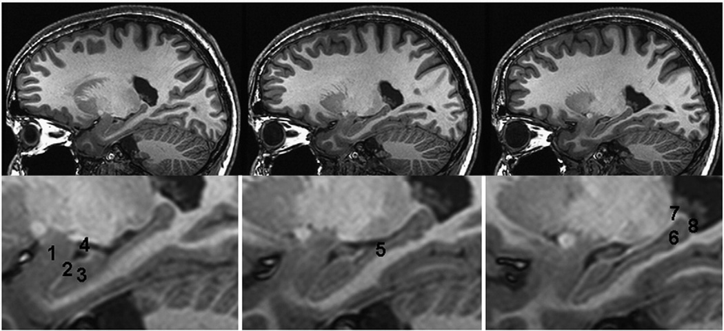
Fig. 1.
Gray and white matter of the hippocampus. Sagittal MR images of the hippocampus (from medial slice–left image to lateral slice–right image) and enlarged views of the hippocampal area (lower row) demonstrating gray matter of the hippocampal head, body and tail, overlaid by white matter of the alveus and fimbria and amygdala. 1=amygdala, 2=alveus, 3=hippocampal head, 4=temporal horn of the lateral ventricle and fimbria, 5=hippocampal body, 6=hippocampal tail, 7=fimbria, 8=alveus. The images displays the average image of three MR images of a young healthy male acquired using a 0.5×0.5×0.5 mm3 resolution after zero filling at 3 T field strength (Gyroscan Intera 3.0 T, Philips, Best, NL). Raw images are a courtesy of Dr. H. Schiffbauer.

