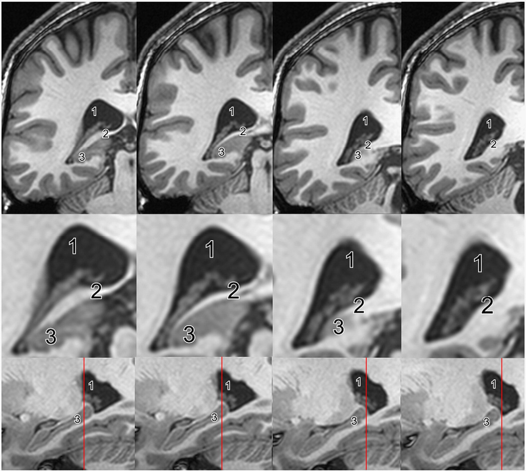
Fig. 3.
Posterior end of the hippocampus. MR images demonstrating the posterior end of the hippocampus. Coronal slices from anterior (left) to posterior (right) and enlarged views of the hippocampal area (middle row). Sagittal slices with the position of the coronal plane indicated by a red line (lower row). While some segmentation protocols stop measuring the hippocampus when the crus of the fornix appears (image two to the left), others follow the hippocampus posteriorly until the hippocampal ovoid gray matter completely disappears (image two to the right). 1=atrium of the lateral ventricle, 2=crus of the fornix, 3=hippocampal tail.

